
Technology Note 200
Receptor Valency Effects in KinExA Measurements
Kinetic Exclusion Assay (KinExA®) experiments determine the free fraction of a receptor in a sample containing receptor, ligand, and receptor-ligand complex. Using this information the concentration of one of the analytes or the Kd of a binding pair can be derived, depending on the experimental conditions. The theoretical models used for the determination assumes 1:1 binding, while in fact most of the time the valency is greater than one. Does this cause an error in the experimental results? This tech note will explore the effect of receptor valency on experimental results, including when and how valency will affect the measurements.
Source of Multivalency Measurement Errors
In any assay that relies on capturing a free binding site (this includes ELISA, SPR, and KinExA), the signal is related to the amount of receptor that is captured. Once captured, a receptor gives the same signal whether it had one, two, or more free binding sites in solution. Therefore, the signal does not directly reflect the number of free binding sites when the receptor is bivalent (or more generally multivalent). Although the signal from a captured receptor is independent of the number of free binding sites, the probability of being captured can be affected by the number of free binding sites.
If the overall capture probability is low, the capture probability converges to being proportional to the number of free binding sites, so that the signal once again is proportional to the free binding sites in the sample. A complete mathematical treatment of this has been published1, concluding that for low capture probabilities, the error can be ignored.
In KinExA measurements the contact of the sample to the solid phase is kept intentionally short, unlike other capture assays. The short contact time is to kinetically prevent competition between the solution ligand and the solid phase ligand for the receptor. While this works well in allowing samples to be measured without perturbing the bound fraction, it also tends to keep the capture probability low, giving a more accurate measurement of free binding sites when using multivalent receptors.
Capture Probability and Measurement Error
The original mathematical treatment of capture probability and measurement error2 only discussed the error in the free binding sites estimated from the measured signal as a function of the overall capture probability. Of more relevancy is the resulting error in Kd measurement. To get a visual representation of the effect on a Kd measurement, a theoretical case of a bivalent receptor (e.g. an antibody) was analyzed at different capture probabilities to generate a graph of calculated Kd values.
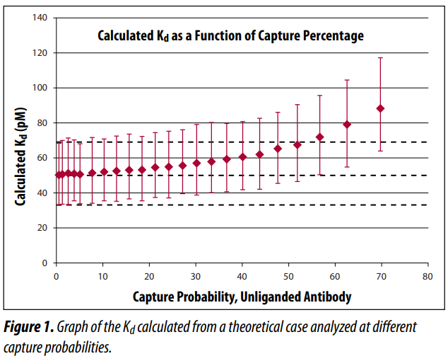
In Figure 1, the X axis is the capture probability for a bivalent receptor. The Y axis is the Kd value expressed in units of picomolar (pM). Each data point is the average of 1000 independent analyses, using 3% random noise and run through the standard KinExA analysis software. The error bars indicate a range encompassing 95% of the individual Kd values.
Conclusions
As long as the capture probability is below 40-50%, the error caused by the bivalency of the receptor is well within the normal measurement error. While the capture probability is not normally measured as part of an analysis, it has been reported in publications that the instrument has a “typical capture efficiency of less than 10%”3 or, more specifically, 1.2% to 5.9%2. Although this is within the desired range, it does vary from system to system. Since the usual goal is to get a highly active solid phase (high capture probability), it may be higher than 50% for some systems. If there is a concern, a simple test can be performed to be sure the capture probability is in a range that is not causing a perceptible measurement error. See How to Guide 250 Capture Percent Test (HG250) for information on how to perform the test.
References
1. Glass T.R. and Winzor D.J. 2014. Confirmation of the validity of the current characterization of immunochemical reactions by kinetic exclusion assay. Anal Biochem 456: 38-42. http://www.ncbi.nlm.nih.gov/pubmed/24751468
2. Ohmura, N., Lackie, S.J., et. al. 2001. An immunoassay for small analytes with theoretical detection limits. Anal Chem 73(14): 3392-9.
3. Blake, R.C., Pavlov, A.R., and Blake, D.A. 1999. Automated kinetic exclusion assays to quantify protein binding interactions in homogeneous solution. Anal Biochem 272: 123-134.
U.S.A. Headquarters

Phone
(208) 345-3400
Emails
Sales: sales@sapidyne.com
Accounts Payable: ap@sapidyne.com
Accounts Receivable: ar@sapidyne.com
Admin: admin@sapidyne.com
Tech Support: support@sapidyne.com
Customer Service: info@sapidyne.com
Address
700 W Diamond St
Boise, Idaho 83705-5258
USA
European Office

Phone
+49 173 242 7044
Emails
Sales: sales@sapidyne.eu
Accounts Payable: ap@sapidyne.eu
Accounts Receivable: ar@sapidyne.eu
Admin: admin@sapidyne.eu
Tech Support: support@sapidyne.eu
Customer Service: info@sapidyne.eu
Address
Feodor-Lynen-Str. 21
30625 Hannover
Germany
Distributors
Japan
Tokyo
+81 (0)3 5625 9711
sales@scrum-net.co.jp
www.scrum-net.co.jp
Korea
Seoul & Daegu City
+82-10-4804-0454
greenics@naver.com
www.greenics.co.kr
China
Beijing Rohe Trading Co., Ltd
Beijing
010-8283-4408
info@loham.cn
www.loham.cn
Singapore
Yiches
+65 6498 0927
sales@yiches.com.sg
www.yiches.com.sg